Surgical AI · VirtaMed · 2022 – now
Ultrasound Anatomy Detection
Finding anatomy in ultrasound, from 60% to 90% accuracy.
- 60% → 90%anatomical structure detection accuracy
- Automaticannotation, straight from the simulation
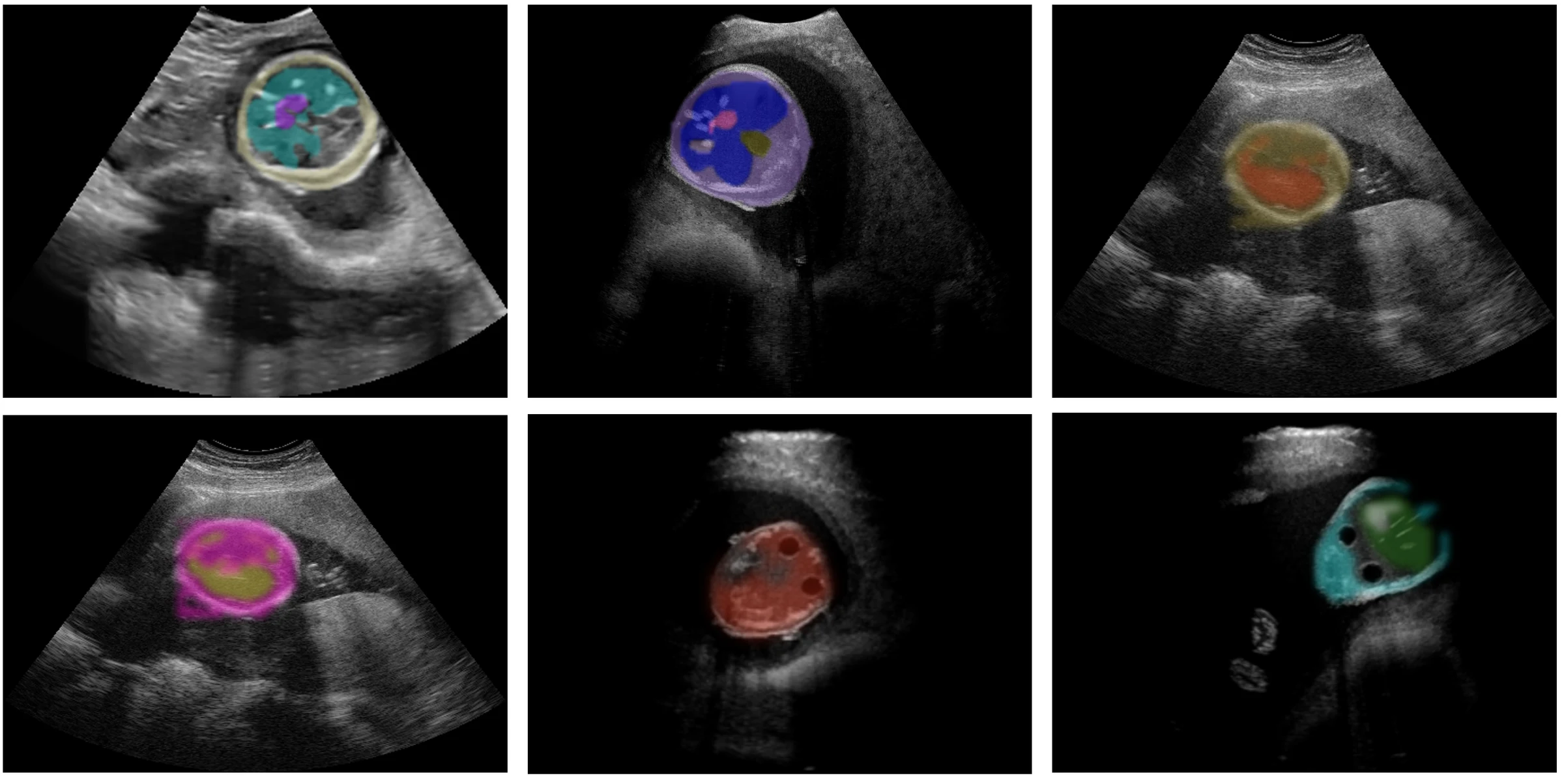
Detecting anatomical structures in ultrasound images with transformer-based detection models, which raised accuracy from 60% to 90%. Shown with a pipeline that produces labelled training images from simulation.
The problem
Ultrasound is hard to read and expensive to label. Every annotated image needs a clinician’s time, and a detector needs a great many of them.
The approach
Let the simulation do the labelling. A simulated ultrasound image is rendered from a 3D model, so the position of every structure in it is already known.
- 3D modelling. The anatomy starts as a 3D model.
- Ray tracing. An ultrasound image is rendered from it.
- Automatic annotation. The labels for each structure are produced together with the image.
- AI on synthetic data. Detection models are trained on the generated set.
- Inference on real data. The trained model is applied to real scans.
Result
With transformer-based detection models, anatomical structure detection in ultrasound images improved from 60% to 90% accuracy.
This is one use of the scalable data-processing pipeline I designed at VirtaMed, which converts 3D simulation output into training-ready datasets for machine-learning and LLM-based models.
Figures
