Research · Amgen Scholars · ETH Zurich · 2018
Alzheimer's Brain Changes with GANs
Showing what Alzheimer's disease does to one specific brain.
- Cambridgepresented at the Amgen Scholars Symposium, 2018
- Deformationmodel instead of an additive one
Amgen Scholars research at ETH Zurich's Computer Vision Lab. A Wasserstein GAN learns a deformation that maps a brain MRI between disease stages, making subject-specific tissue loss visible.
The problem
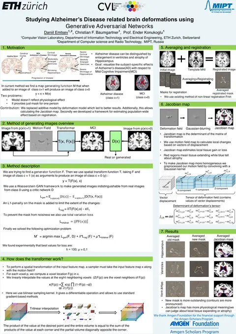
Alzheimer’s disease shows up in MRI as enlarged ventricles and a shrinking hippocampus. Group statistics describe the average patient. The goal here was to visualise the effect for one individual: what separates this brain at the mild-cognitive-impairment stage from the same brain with Alzheimer’s?
The idea
An earlier method learned an additive map: a generator produced a difference image that, added to a scan of one class, yields a scan of the other. That does not reflect physiology, because disease does not add intensity, it moves tissue.
I replaced the additive model with a deformation model. The generator predicts a motion field, a spatial transformer warps the input image with it, and a Wasserstein critic judges whether the result is indistinguishable from real scans of the target class. An L1 penalty limits the extent of the change and a total-variation term keeps the field smooth.
What it gives
- Better results than the additive baseline.
- A Jacobian map computed from the motion field, which estimates local tissue gain or loss at each voxel.
- A framework for population-wide effects, by registering individual maps to a common template and averaging.
With Christian F. Baumgartner and Prof. Ender Konukoglu at ETH Zurich’s Computer Vision Laboratory. Presented at the Cambridge Amgen Scholars Symposium in 2018.